
Medical & Scientific Illustration
Specialized medical and scientific illustration
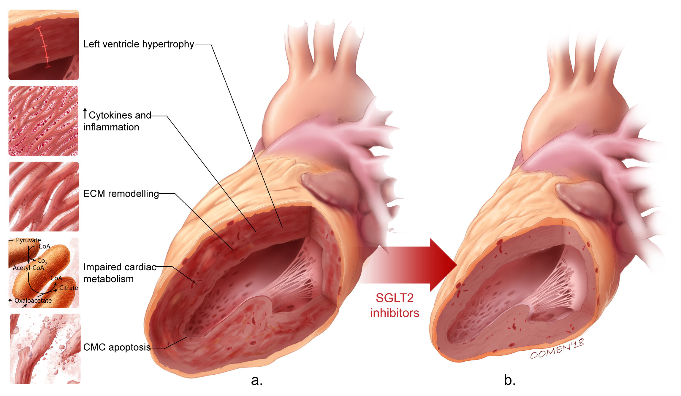
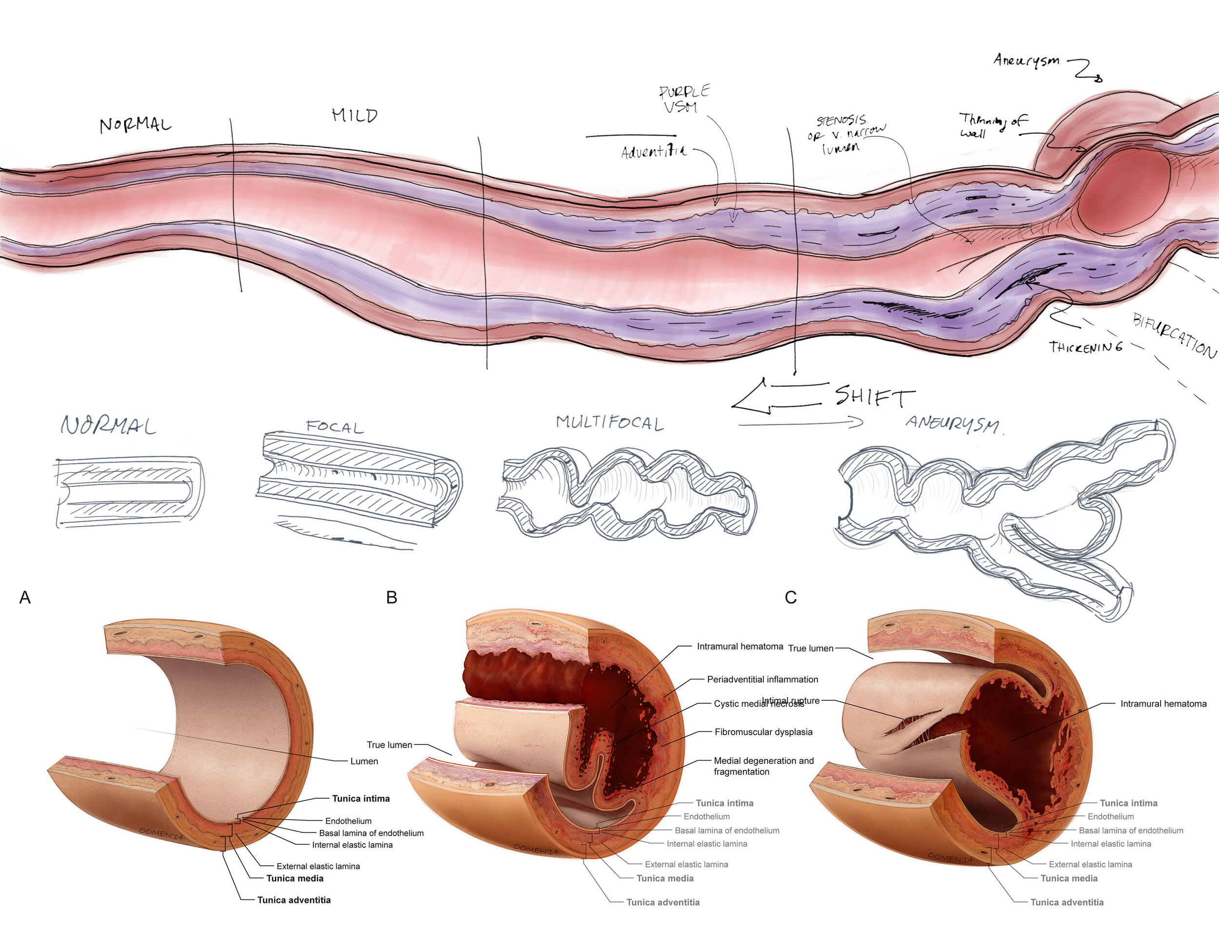
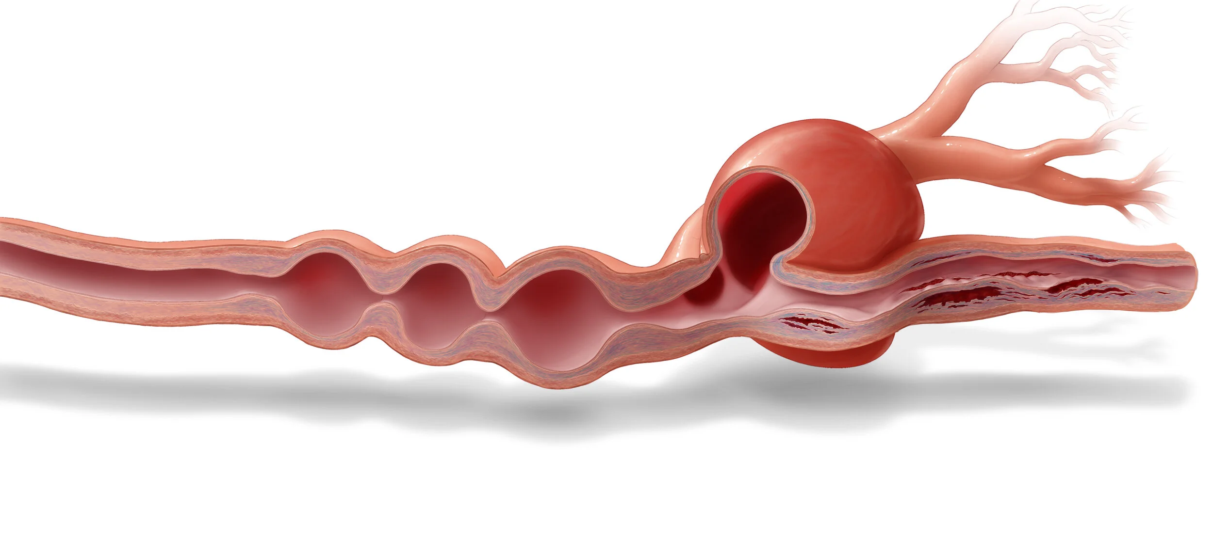
Featured Illustration
This series of images describes the pathology and histology associated with PSCAD, or Pregnancy-related Spontaneous Coronary Artery Disease. The illustrations combined current data about histological changes to the arterial wall with broader, more typical features associated with atherosclerotic plaque development to create compact, detailed and very effective figures. These images were originally used for publication.
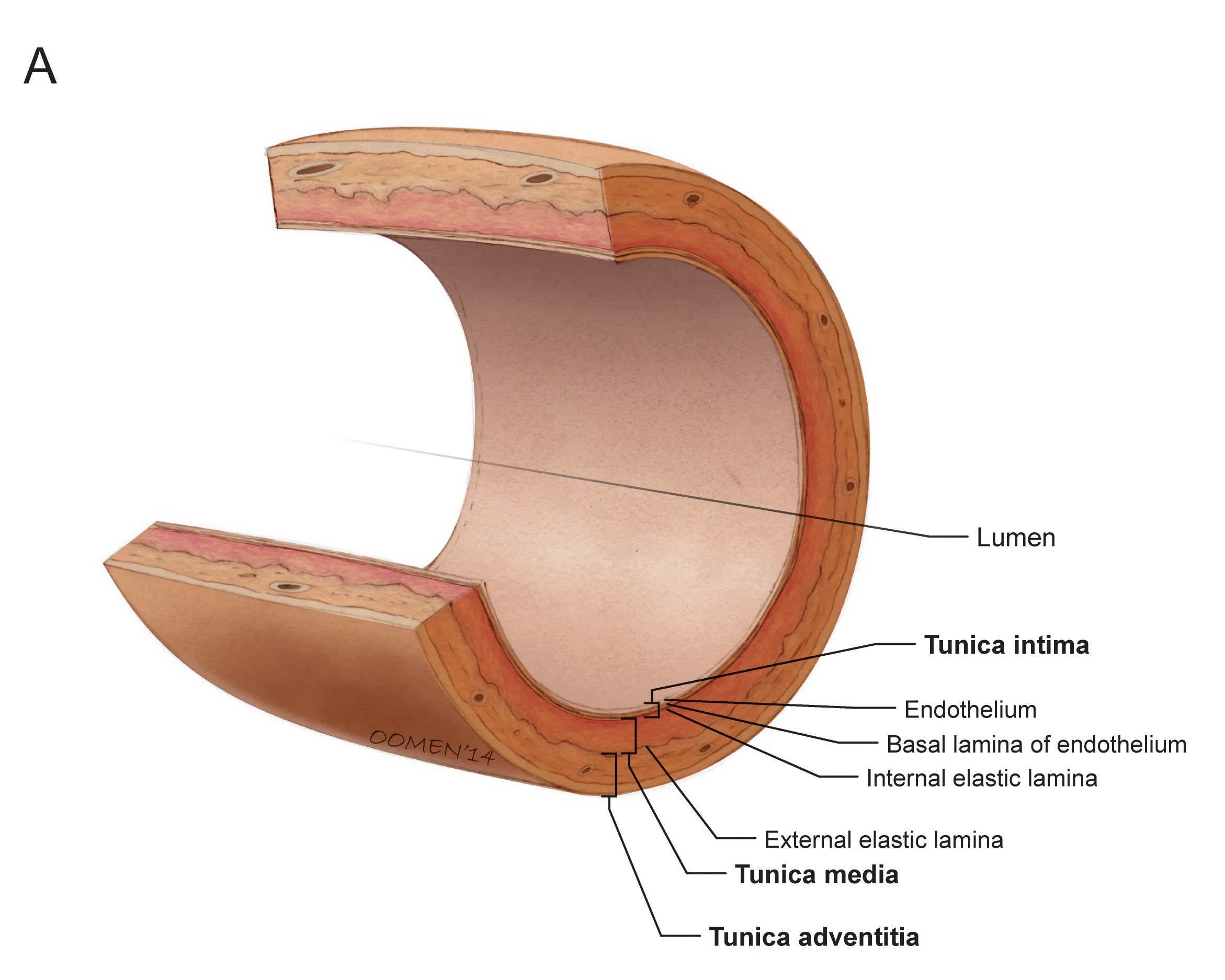
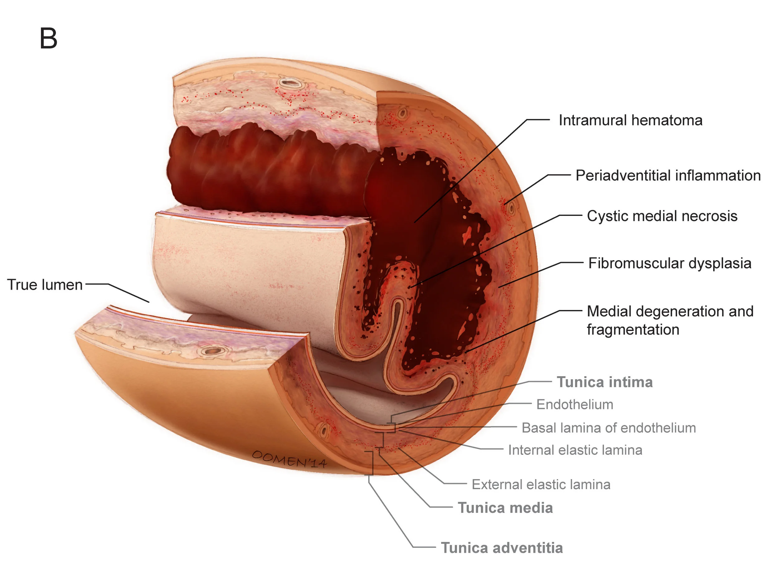
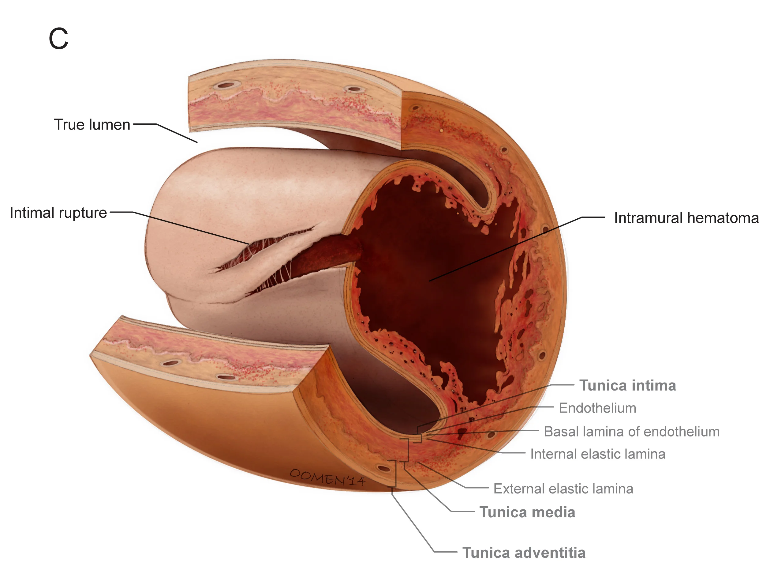
Image copyright 2014, Glen Oomen & Dr. Jacqueline Saw, Vancouver General Hospital / University of British Columbia
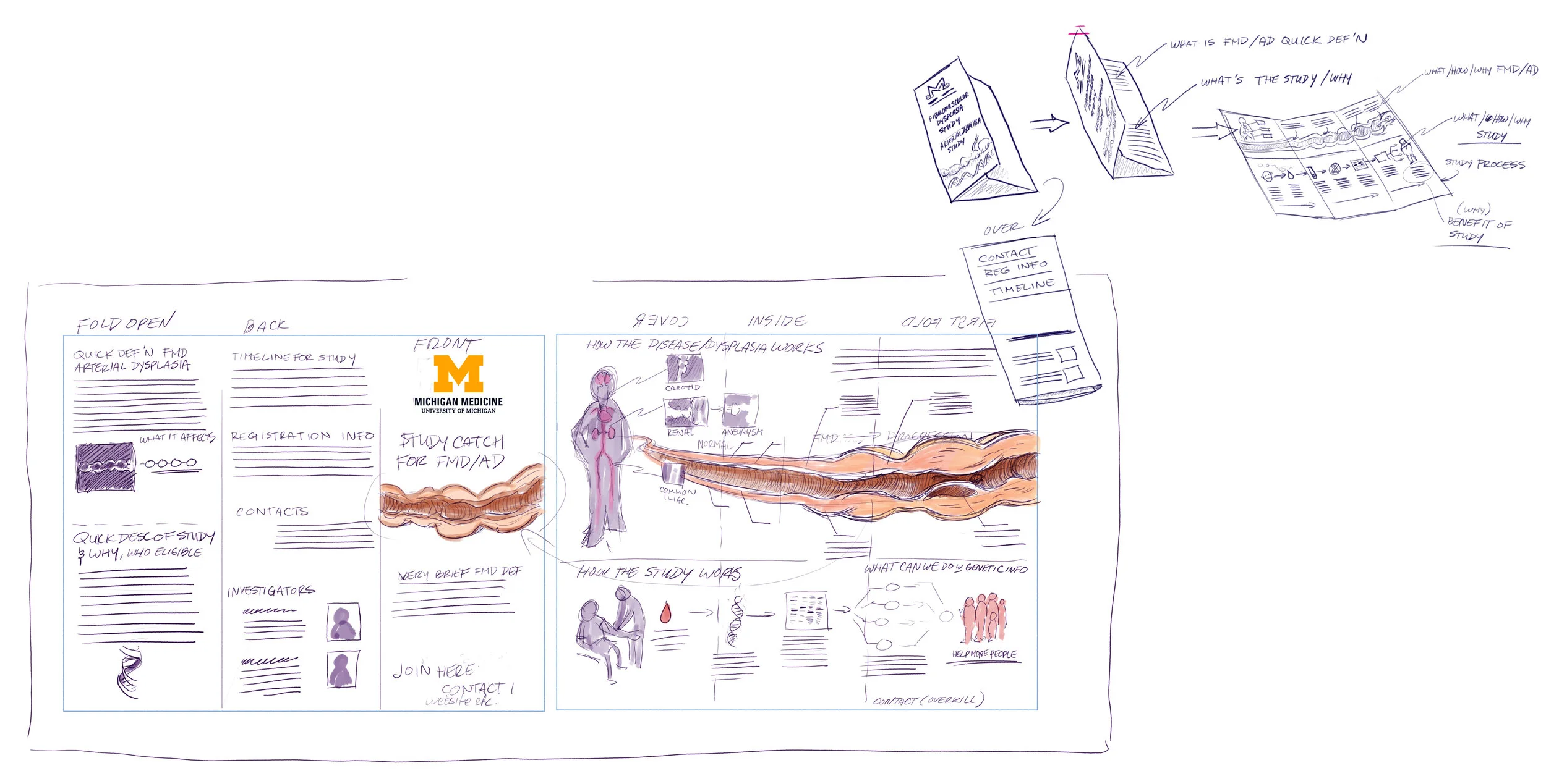
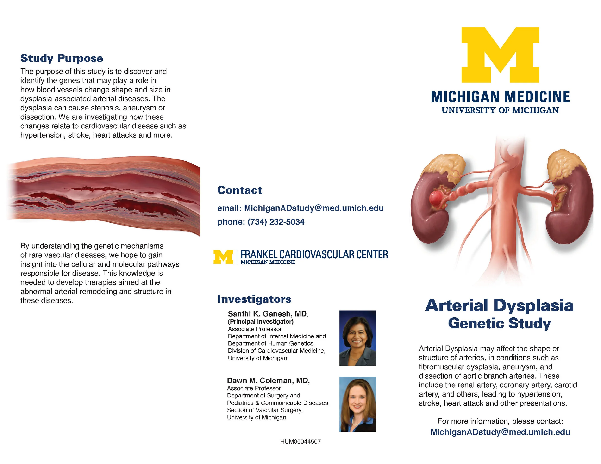
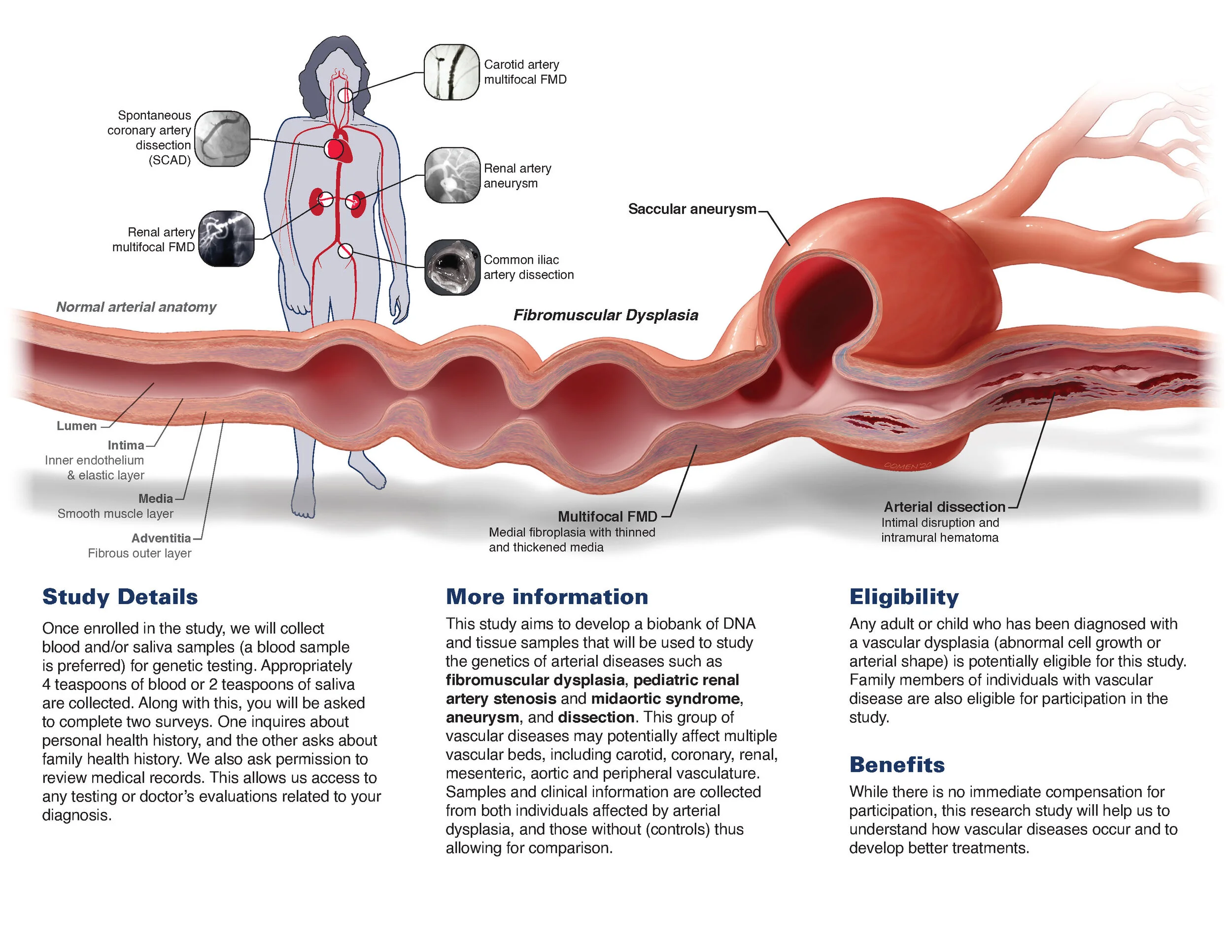
Illustration demonstrating the visualization of the optic nerve sheath as a means of assessing intracranial pressure. In this procedure a slight dilation of the optic nerve sheath, an extension of the dura mater, is visualized and used as a fast, non-invasive means of assessing how much sub-dural or intracranial pressure is present.
Illustration describing how arrays of microneedles can be used for bio-sensing and drug delivery. The needles themselves can be vary in shape to serve as anchors within the skin, preventing removal, or be configured to slowly dissolve, releasing a drug as they do or physically releasing from the tissue after a prescribed period of time.
This is one image from a larger series describing a technique for using tube retractors and guidance systems to access deep brain lesions. This image shows one of the final steps in the sequence, placing a larger diameter tube.
Neurosurgical journal illustration showing endoscopic view of the Superior Hypophyseal Artery (SHA) complex in relation to other critical features and landmarks during transphenoidal access.
Orthopedic journal illustration describing the general anatomy of the sternum and vascular considerations around midline sternotomy.
Illustration showing suturing of linea alba tendonous abdominal fascia, post midline sternotomy.
Final image in sequence for urology journal article describing a technique for creating a neobladder from distal jejunum, showing the pressure testing of the final, constructed bladder.
Journal of Biological Chemistry cover for study describing a gene deficiency that predisposed cancer patients to irreparable cardiac cell death when treated with doxirubicin. The authors, cardiac surgeons, advocated for genetic testing prior to chemotherapy to ensure the safe use of of the chemotherapuetic
This illustration is one of a series describing a technique for cerebral protection while repairing ascending aortic aneurysm with dacron graft. The series specifically shows the placement of extracorporeal line placement to maximize blood flow to the head and brain.
This series of illustrations compares two different techniques for addressing mitral valve prolapse. In the top series, the prolapsing leaflet is resected. In the lower series artificial chordae are sutured in place, leaving a larger diameter final valve.
Illustration series done for New England Journal of Medicine article. This series relates different morphologies of Bicuspid Aortic Valve to the shear forces inflicted upon the aortic wall by their corresponding ejection fractions.
Surgical journal illustration showing three variations in aortic root replacement and repair.
One illustration from a large series describing different techniques for grafting small diameter vessels for coronary artery bypass. This image shows a piggyback graft and a Heartstring device being used.
Illustration showing the course of the posterior cutaneous nerve of the forearm and how it relates to lateral epicondylitis.
Sodium Glucose cotransporters are responsible for the resorption of sodium and glucose in the kidneys. SGLT2 Inhibitors thus decrease the resorption of both solutes, decreasing both blood glucose and sodium as they two are excreted. This allows the myocardium to recover both structurally and metabolically.
Project Workflow
These are some images showing the working process of a project and how it progresses. These are from a recent project completed for the University of Michigan, developing a fairly simple information pamphlet that would be distributed to potential practitioners, study recruiters and patients to educate them on a rarely discussed disease that primarily occurs in renal arteries. It’s not the most sophisticated or high tech of communication mediums, but it’s still possibly the most effective and cost effective: you can take it with you, it won’t run out of batteries, you don’t have to turn it on and you can still take notes on it - that’s paper. The real trick is in how you present the information. Even with the constraints of one piece of paper and a budget, creativity and skill can still produce something beautiful.
Everything begins with sketches. Sometimes, the rougher the better so that ideas don’t become too permanent too early. This rough sketch showed the general layout of information, what was text, what was image, and the overall balance background whitespace and figurative information.
Rough sketches also helped the team refine the generalized anatomy and pathology of the condition so that it could all be shown in one place, progressing from normal and healthy at the left to very pronounced and diseased at the right. Elements were drawn from previous work created for research colleagues that captured much of the shared histology and pathology.
This project had one major “hero” image, the cross section of progressing pathology that could be used by patients and practitioners to understand or explain the disease respectively. But it can’t do all the lifting, and the repetition can diminish its value. Since the disease occurs most often the renal arteries, and we hadn’t yet allotted some space to show and explain that, we decided to create a nice image of kidneys and aorta and diseased renal arteries for the cover to put the condition in situ.
The “hero” image gets completed while the rest of the text and image information is settled on. This image shows the disease from “normal” and healthy at the left to a section where there are ring-like regions with higher expression of vascular smooth muscle, creating strictures. Hemodynamically, these strictures predispose the artery to outpouching and aneurysm, particularly where the vessels branch and bifurcate. Lastly, the imbalance in tissue and hemodynamics created regions of endothelial dysruption and mural bleeding.
The outside of the brochure has just enough information to hook the viewer, but not enough to horrify them. Medicine is full of things that people don’t understand, so you need to by mindful of what information gets presented when in their journey. In this case, the kidneys are the front since this is really initially diagnosed or seen as a renal problem. Then the nuts and bolts and stuff that’s not so nice to look at (unless you’re a medical illustrator or physician who’s into this stuff) on the inside. This way, nobody is too embarrassed or disgusted to pick up information that’s really there to help them.
Then everything gets put together. It’s always satisfying to see each piece of the information - visual, text, even white space - all working as one to move the reader about the page in the right ways
This neurosurgical image describes the burr hole placement and an endoscopic approach angle for accessing 3rd ventricle tumors and lesions. In this image, there is pineal tumor on the supero-posterior wall of the 3rd ventricle, but the approach also enables access to mammillary bodies.